Inside the pumpkin
by Rob Graham
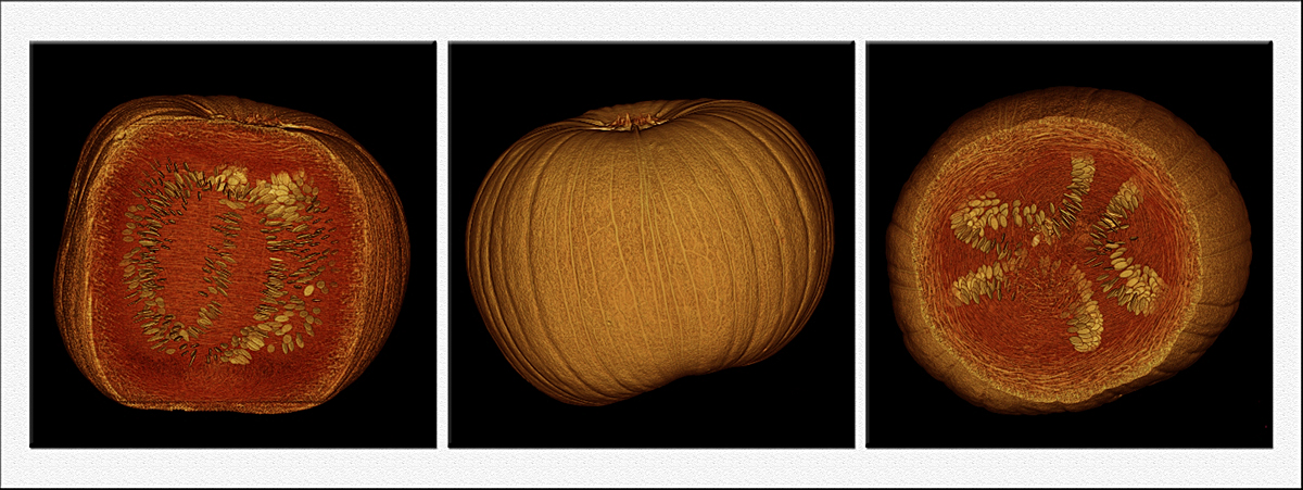
Gary Yeoh's work with flowers and a mammography unit inspired me to attempt the same sort of work. I didn't have much luck, but also haven't spent the time to master it. Instead, I took a different approach to combining radiography and flora. For this series, I scanned a pumpkin with a CAT scan unit at work, then reconstructed the data to create these images. Using the reconned data, I then cut away part of the pumpkin to reveal a side view and then part of the top of the pumpkin to reveal a view from the top. One issue I had when working with these is they are only 512x512 natively, so I had to enlarge them. The seeds especially show the jaggies inherent to a low pixel matrix. I found the patterns of the seeds to be quite interesting. The recon software actually imparted a slight orange cast to the data, which I then enhanced in CS5.
Moderators, if this is to far fetched for this forum, please don't hesitate to delete or move the image.
Siemens Somatom Sensation 64, 495 slices at 0.6mm each, Aquarius workstation for reconstructions. C&C always welcome and appreciated.

Comments
9 commentsBill, thanks for letting this stay, and if you find a good deal on a CT or X-ray machine, let me know :).
Ian, a modern CAT scan machine creates a volume of info as it scans, which can then be cut into individual slices. In this case, I chose a 0.6mm thickness per slice, as it is the thinnest our machine can create. Those 495 slices were then sent to the Aquarius workstation, which essentially stacked the images to recreate the volume of info, as well as performed surface rendering. I then manipulated that image to create the above images. I wish I had CS5 Extended, as I think it handles dicom files and can do 3D rendering. I am including a single 0.6mm slice as a reference for how the original data looks; the image is from the middle of the pumpkin.
Tonia, I am lucky to have an open-minded supervisor :). CT is my favorite modality, though I also regularly work in Nuclear Medicine and diagnostic x-ray.
Take a peek in the Photo Art gallery for a different look at this subject :).
Sorry for coming to the party late. Was just too busy in the last few weeks to even peep in.
W
I was going to try the CT scan at some stage but you have beaten me to it. At least now I know it will work and there is no need to do the proof of concept work. What is most interesting for me is the fact you could use the software to render the color into this grayscale image. Your photoart image is way out in the energy realm!
Look forward to seeing more of your Xray images.